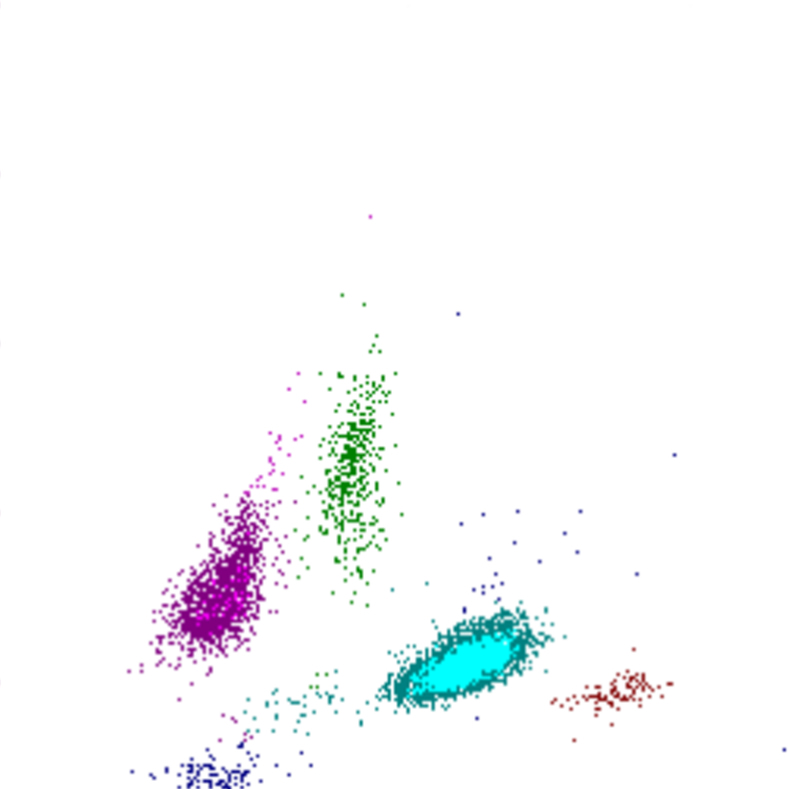
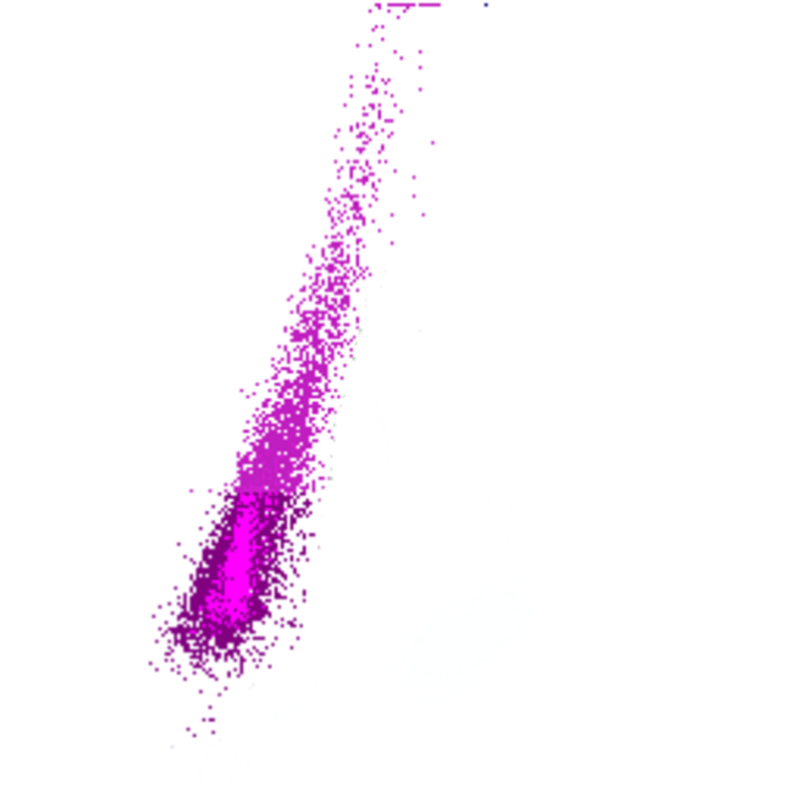
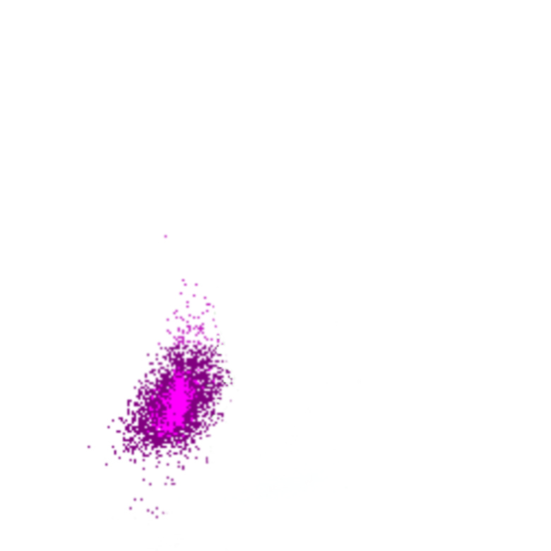

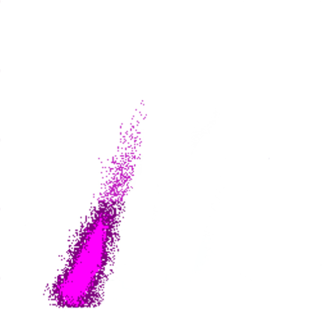
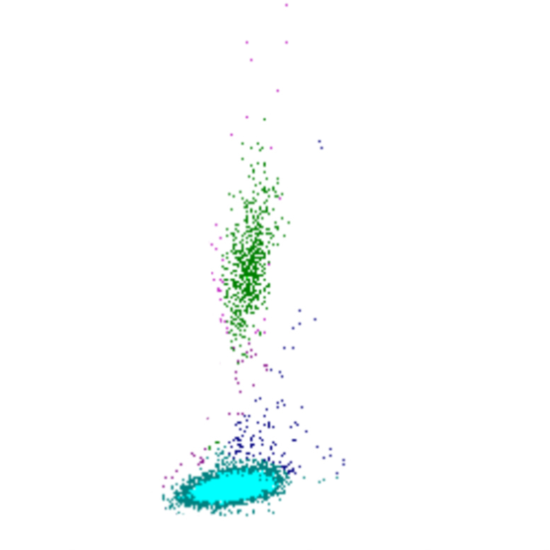
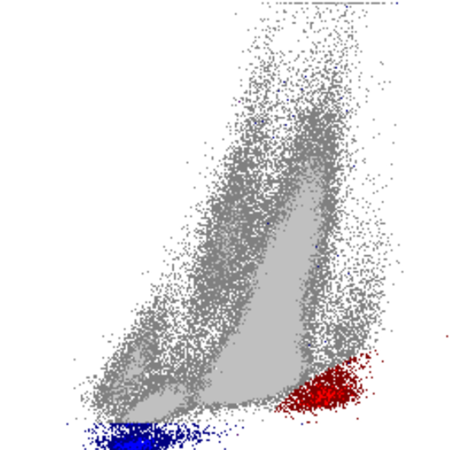
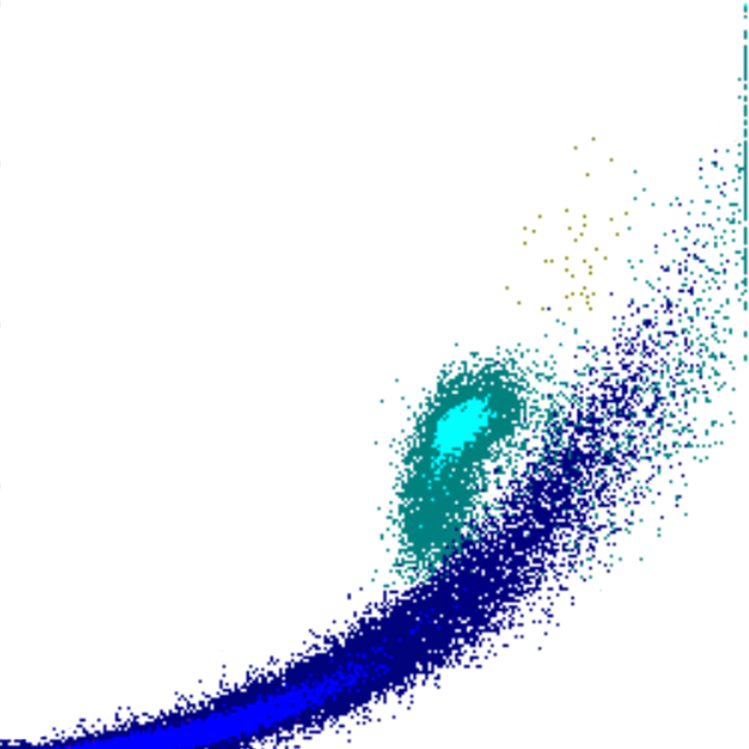
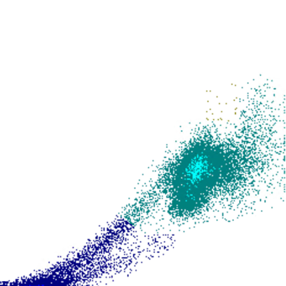

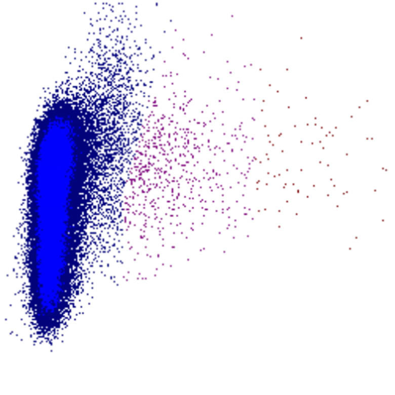
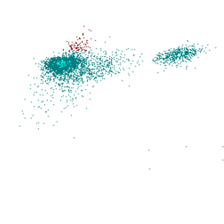
WDF - Fibrin
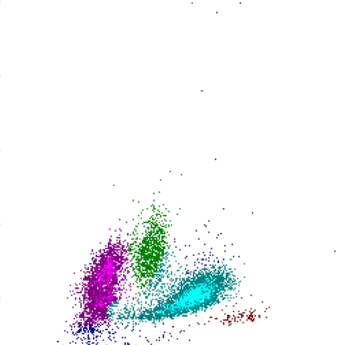
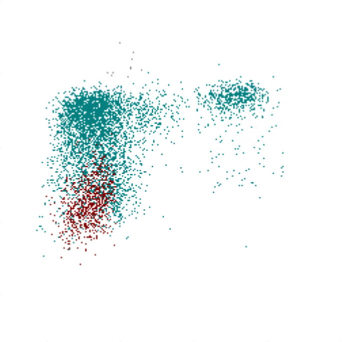
SFL
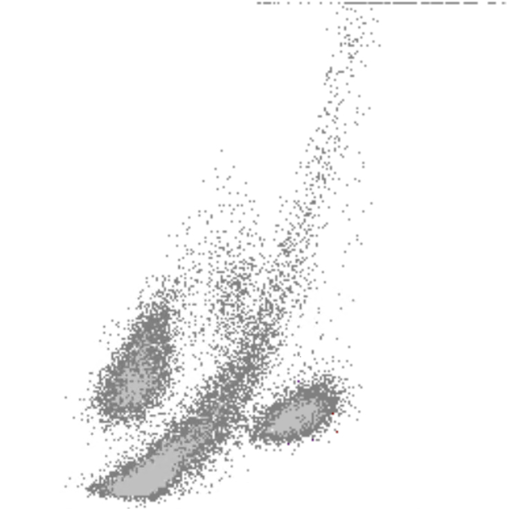
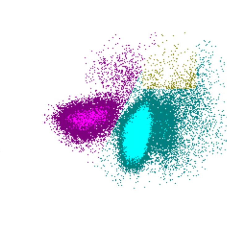
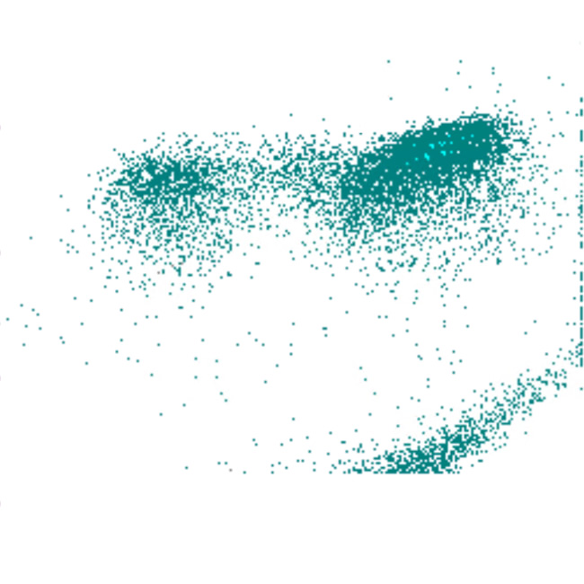
SSC
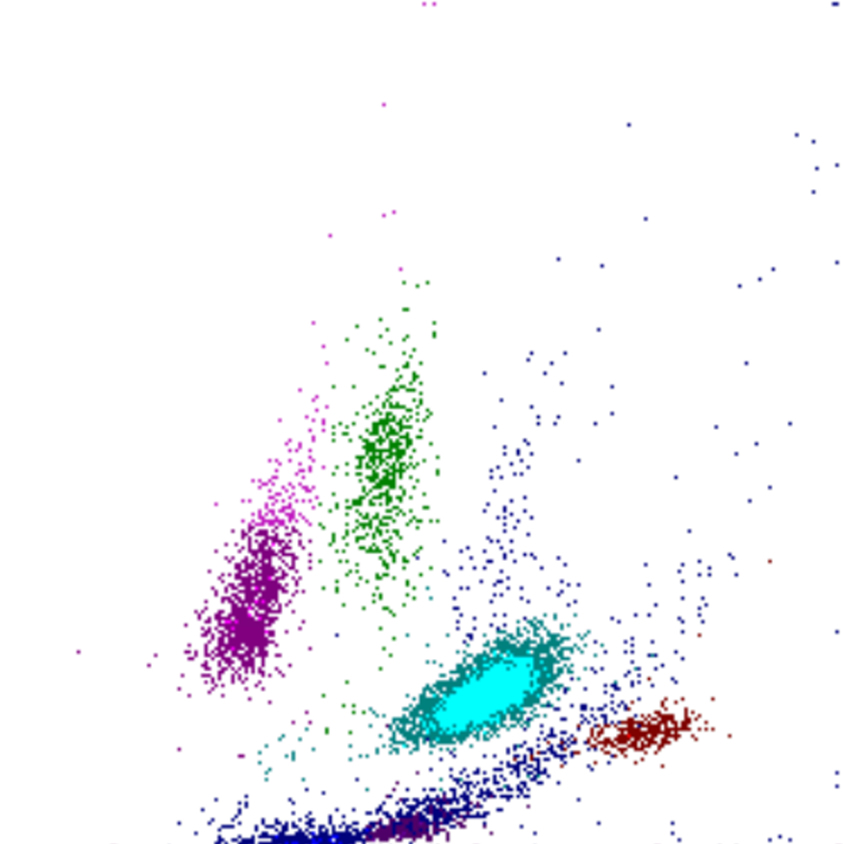
Lymphocytes
Monocytes
Neutrophils
Eosinophils
Basophils
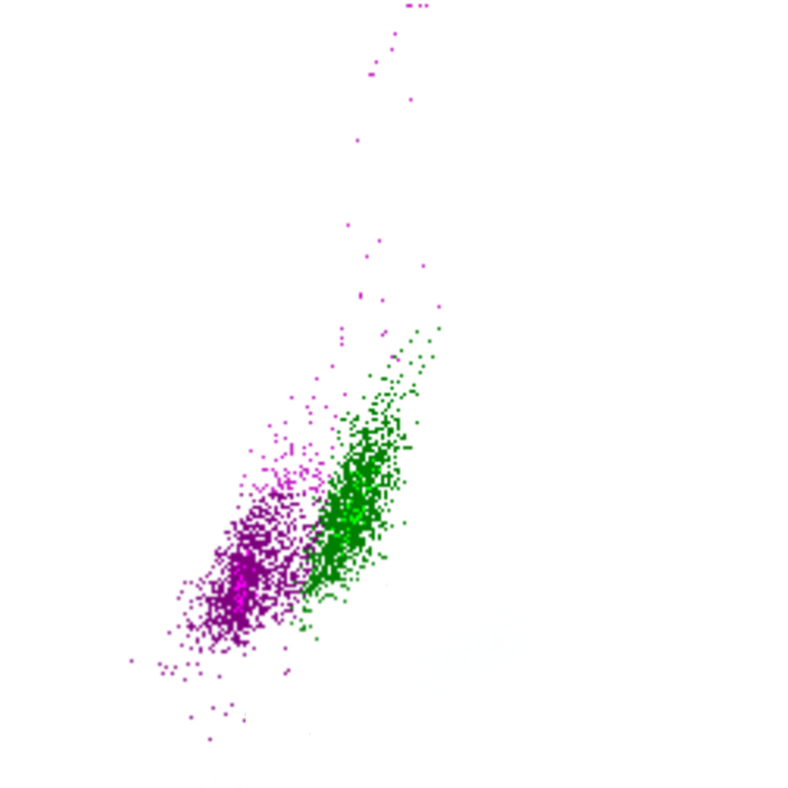
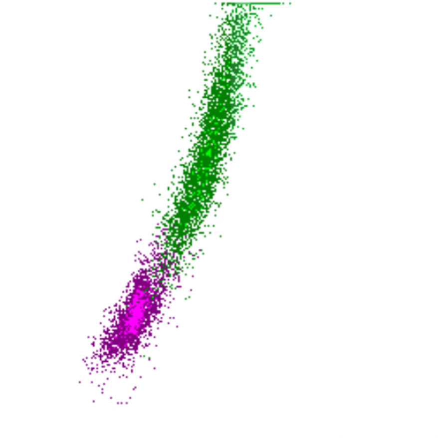
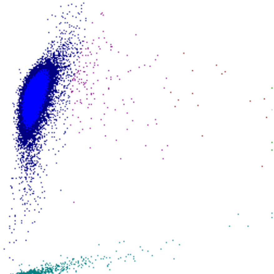
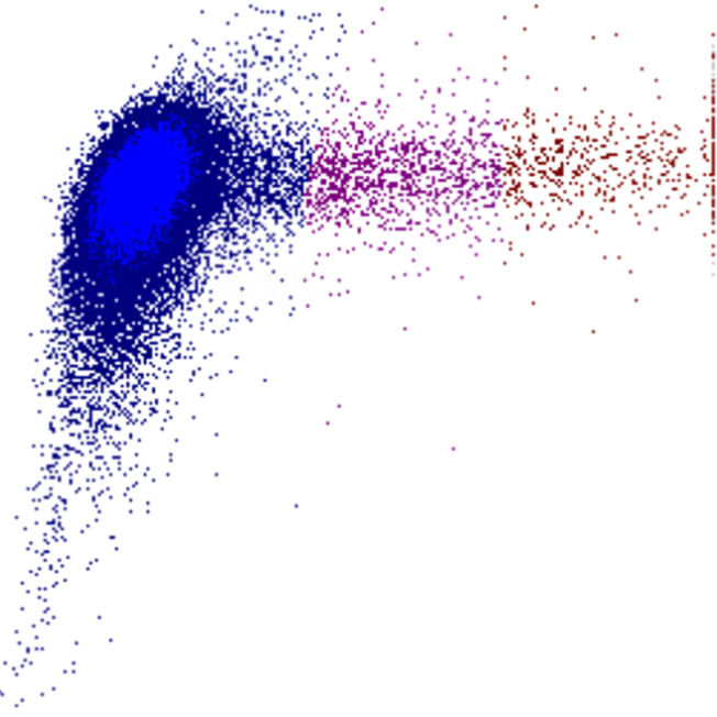
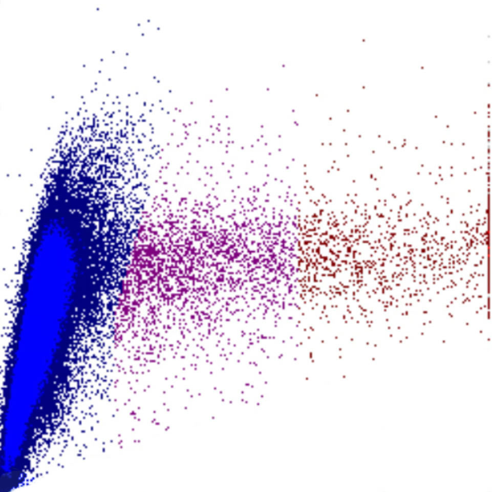
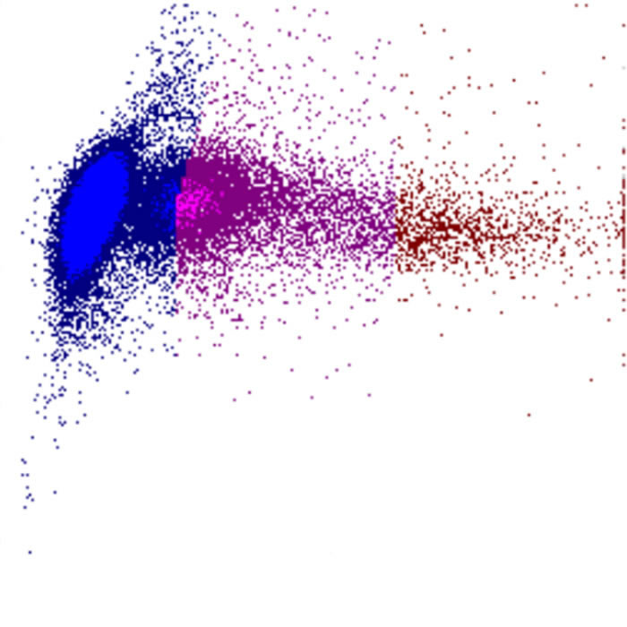
In this sample, the WDF plot shows a blue tinge extending from the debris region between the neutrophilic and eosinophilic granulocytes . This extra population is caused by the presence of fibrin filaments in the sample.
In the WDF channel, fibrin filaments may partially fluorescently stain and generate scatter, becoming visible as an elongated, diffusely bounded population. This represents a pre-analytical artifact, usually due to insufficient mixing, delayed processing or partial coagulation of the sample.
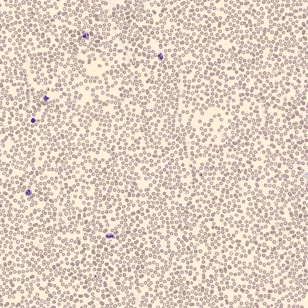
Possible morphology